


Diagnosis of Tinnitus
When a patient appears with tinnitus, the clinician’s primary priority is to determine if the source is treatable or potentially harmful.
HISTORY AND PHYSICAL EXAMINATION
Tinnitus is diagnosed mostly by a focused, detailed history and physical examination, which distinguishes between benign and harmful causes and determines appropriate treatment options. Initial triage is determining which aspects of the patient’s tinnitus require immediate evaluation: pulsatile, coupled with neurologic abnormalities, asymmetric or unilateral symptoms, and asymmetric hearing loss. Although it is uncommon, pulsatile tinnitus should be investigated for underlying cardiovascular illness and cerebral vascular abnormalities (e.g., vascular tumor), elevated intracranial pressure, or malignancy. The four most useful history components for evaluating and managing tinnitus are chronicity, location (unilateral or bilateral), any concomitant hearing alterations, and impact (bothersome or not bothersome). The patient should be asked about any accompanying symptoms, such as sleep and mood disturbances, as well as cognitive impairments, which have a negative impact on quality of life and may improve with treatment. The Tinnitus Handicap Inventory and Tinnitus Questionnaire are the two most commonly used measures in studies to assess the impact of tinnitus on daily life. The Hearing and Tinnitus Survey, a recently validated instrument, is a simple questionnaire that can distinguish between the burden of tinnitus and hearing loss, which may be valuable to clinicians. The physical exam should cover the head, eyes, ears, neck, neurologic system (with a focus on cranial nerves and cerebellar function), and cardiovascular system.
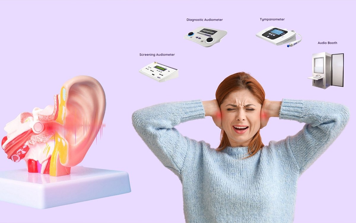
AUDIOLOGY
A full audiologic evaluation is recommended for each patient with tinnitus, regardless of duration or characteristics. The American Academy of Otolaryngology-Head and Neck Surgery (AAO-HNS) recommendation suggests that patients with chronic tinnitus (lasting six months or more), unilateral tinnitus, or any reported hearing changes be referred for full audiologic testing within four weeks. Audiometry can detect the presence, severity, type (conductive, sensorineural, or mixed), and asymmetry of hearing loss. Patients who arrive with tinnitus coupled with sudden SNHL should receive prompt audiometry as indicated by the AAO-HNS recommendations, as this necessitates immediate treatment with intratympanic corticosteroids. If audiometry is not available immediately, it should be completed within two weeks.
IMAGING AND OTHER DIAGNOSTIC TESTING
The AAO-HNS and European guidelines urge imaging investigations for patients with unilateral, pulsatile tinnitus, asymmetric hearing loss, or focal neurologic abnormalities. This recommendation is based on observational studies that assessed the high cost, risk of injury, and low yield of these studies in addition to the previously indicated findings on history and physical examination. The suggested imaging test for asymmetric or unilateral, nonpulsatile tinnitus is magnetic resonance imaging (MRI) of the head and auditory canal with and without contrast medium. This assessment is the ideal test for detecting vestibular schwannoma, the most frequent cerebellopontine angle lesion, and tumors in the auditory pathway. If MRI with contrast media is not available or is contraindicated, non-contrast MRI, the auditory brainstem response test (ABR), and computed tomography (CT) are alternatives. Non-contrast MRI of the brain is comparable in sensitivity to detecting vestibular schwannoma but may be less appropriate for identifying other causes of tinnitus. The auditory brainstem response test can also be used to screen for vestibular schwannoma, and it is highly sensitive for lesions bigger than 1 cm but not for smaller ones. CT scans can detect vascular or osseous diseases, although they may miss tiny masses that cause symptoms. The recommended investigation is a temporal-bone CT scan without contrast medium or a head and neck CT angiography. A temporal-bone CT without contrast medium looks for paragangliomas or adenomatous middle ear tumors. CT angiography of the head and neck looks for underlying vascular issues such as carotid stenosis, dural arteriovenous fistulas, or evidence of spontaneous or idiopathic intracranial hypertension, among other reasons for pulsatile tinnitus. If iodinated or gadolinium contrast media are not available, non-contrast MRI and magnetic resonance angiography can still be employed to examine vascular processes. Ultrasonography can detect extracranial carotid stenosis, but positive results are usually followed by CT angiography or magnetic resonance angiography. Laboratory test findings are unlikely to reveal the source of tinnitus; hence, testing should be directed by clinical suspicion for particular contributory disorders. Previous studies linking tinnitus to thyroid disease, vitamin B12 insufficiency, and diabetes mellitus were based on very low-quality evidence, so routine testing for such disease entities is not recommended. If the doctor is concerned about syphilis or Lyme disease, low-quality evidence indicates that suitable serologies may be beneficial; nonetheless, the incidence is modest.
Prevention of Tinnitus
Tinnitus prevention is significantly connected with SNHL; thus, strategies to reduce occupational and recreational noise exposure may be useful. Earplugs and earmuffs have been demonstrated to help decrease noise-induced hearing loss. Caution with potentially ototoxic drugs, as well as monitoring during usage, may assist in avoiding or treating tinnitus.
TREATMENTS TO AVOID
Benzodiazepines are not advised for treating tinnitus. Benzodiazepines, as a class, have uneven effects, and their usage is associated with a significant risk of side consequences and misuse. Anticonvulsants, such as gabapentin (Neurontin), carbamazepine (Tegretol), lamotrigine (Lamictal), and acamprosate (Campral), have been tested on a modest scale and shown to be unsuccessful in the treatment of tinnitus, with a high rate of side effects (18%). There is no evidence that Ginkgo biloba helps patients with primary tinnitus. Nitrous oxide was likewise determined to be an unsuccessful therapy. The use of repetitive transcranial magnetic stimulation in the treatment of tinnitus is debatable. The most current research on repetitive transcranial magnetic stimulation in the treatment of tinnitus has sparked discussion. According to the most recent research, repetitive transcranial magnetic stimulation is useless and should not be used to cure tinnitus. Electrical stimulation, particularly transcranial direct current stimulation, is ineffective and therefore not recommended. There is insufficient evidence for or against the use of the following treatments for the treatment of tinnitus: acupuncture; electrical stimulation in the form of transcutaneous electrical nerve stimulation; bimodal stimulation, a combination of auditory and somatosensory (electrical) stimulation; hyperbaric oxygen; and a surgical procedure known as microvascular decompression of the cranial nerve VIII (vestibulocochlear).